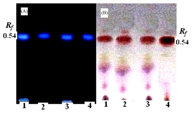
Fig. 6.
TLC of the TTX fraction from different organs of A. stellatus, Lane 1: Muscle, Lane 2: Ovary and Lane 3: Liver, (A) After development, toxin samples were heated for 10 min and visualized under UV light (365 nm); and (B) after development, toxin samples were sprayed with 0.3% of ninhydrin solution.

