FIGURE 1.

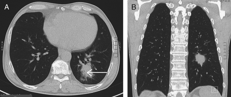
A, B, CT shows an irregular opacity in the left lower lobe. This opacity contains an airbronchogram (arrow) and is surrounded by a ground-glass halo.

A, B, CT shows an irregular opacity in the left lower lobe. This opacity contains an airbronchogram (arrow) and is surrounded by a ground-glass halo.