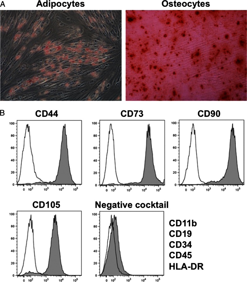
FIGURE 2.

Characterization of cultured hAMSCs. A, Multipotency of hAMSCs. Differentiation into adipocytes was confirmed by the existence of lipid vesicles stained with oil red O (left). Differentiation into osteocytes was confirmed by the existence of mineral nodule deposition stained with Alizarin red S (right). B, Flow cytometry of hAMSCs. The negative cocktail contained antibodies against CD11b, CD19, CD34, CD45, and HLA-DR. Closed areas indicate staining with a specific antibody, whereas open areas represent staining with isotype control antibodies.
