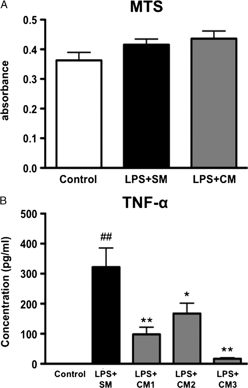
FIGURE 6.

Effect of hAMSC-CM on the activation of Kupffer cells in vitro, (A) Primary Kupffer cells were treated with 100 ng/ml of LPS in standard medium (SM) or hAMSC-CM for 4 h. The number of cells was evaluated by MTS assay. (B) Primary Kupffer cells were treated with 100 ng/ml of LPS in standard medium (SM) or hAMSC-CM for 4 h. CM was obtained from hAMSCs of 3 different donors (CM1, CM2, and CM3). Secretion of TNF-α from the Kupffer cells was measured by ELISA. The values were the mean ± SEM. ##P < 0.01 versus Control. *P < 0.05, **P < 0.01 versus LPS + SM. ELISA indicates enzyme-linked immunosorbent assay; MTS, 3-(4,5-dimethylthiazol-2-yl)-5-(3-carboxymethoxyphenyl)-2-(4-sulfophenyl)-2H-tetrazolium; CM, conditioned medium.
