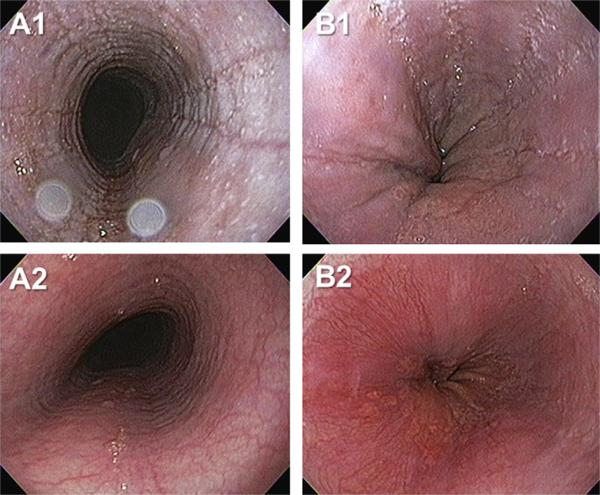
Fig. 1.
Endoscopically identified esophageal features of eosinophilic esophagitis. Images A1 and B1 depict edema, rings, exudates and furrows of the esophagus from two patients (A, B). Images A2 and B2 depict the corresponding images from the same patients following six weeks of therapy with swallowed, topical fluticasone. Interval improvement in edema, exudate and furrows is evident.

