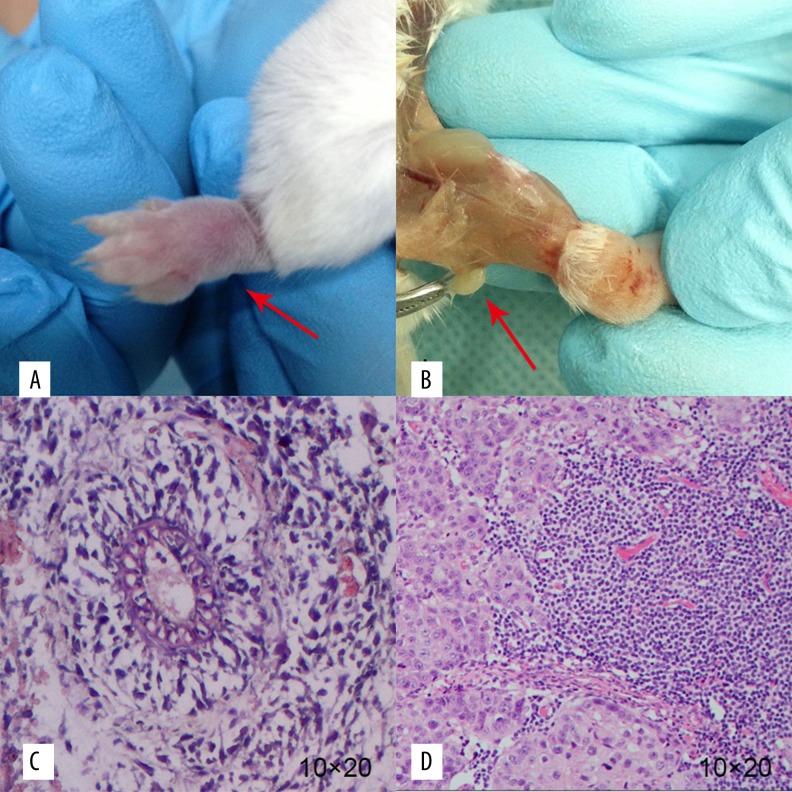
Figure 5.
The general samples and HE staining of transplanted tumor and metastatic lymph node. The red arrows show the general samples of the transplanted tumor and the metastatic lymph node, respectively (A, B). Photograph C and D show the HE staining (10×20) of the transplanted tumor and the metastatic lymph node, respectively. We found cancer cells of different sizes and shapes. Such cells were deformed, mostly in the shape of a nest, glandular tube, or in disorder (C, D).

