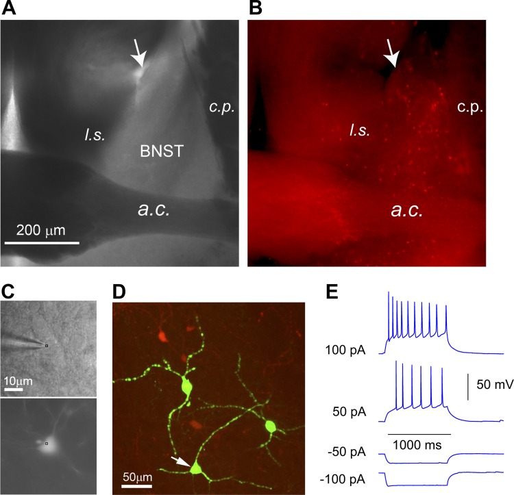
Fig. 1.
Genetic label guided recordings of CRH+ neurons in the BNST slices. To genetically label CRH-expressing neurons, CRH-Cre mice are crossed to the Ai9 tdTomato reporter mice to produce CRH-Cre; Ai9 mice, which express red fluorescent proteins (tdTomato) in CRH-expressing neurons. A and B: bright-field (A) and epi-fluorescent (B) images of a CRH-Cre; Ai9 mouse brain slice show the distribution of CRH-expressing neurons in the BNST. The anterior dorsal BNST (adBNST) is anatomically enclosed by the lateral ventricle (see the arrow), lateral septum (l.s.), caudate putamen (c.p.), and anterior commissure (a.c.). C: CRH+ neurons expressing tdTomato fluorescent proteins (bottom) are targeted for recordings (top) under a high-power objective. D: post hoc visualization of recorded neuronal morphology via intracellular biocytin staining (green). The arrowhead points to the recorded CRH neuron shown in C. E: most CRH+ neurons recorded from anterior dorsolateral BNST show a regular action potential firing phenotype.

