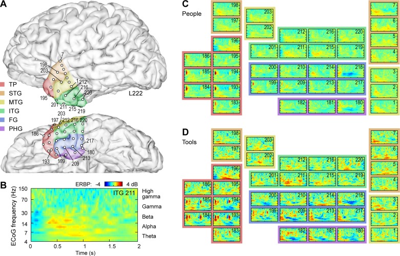
Fig. 3.
Electrocorticographic (ECoG) activity within left anterior temporal lobe (ATL) in a representative subject (L222) during naming tasks. A: anatomical reconstruction of recording site locations within the left ATL. Colors represent the 6 regions of interest [temporal pole (TP); superior temporal gyrus (STG); middle temporal gyrus (MTG); inferior temporal gyrus (ITG); fusiform gyrus (FG); and parahippocampal gyrus (PG)]. B: time-frequency analysis of ECoG activity during naming of U.S. presidents recorded from a representative site (contact no. 211, ITG). C and D: responses from all ATL sites during people and tool naming task, respectively. Each event-related band power (ERBP) time-frequency plot is depicted using the axes as those shown in B.

