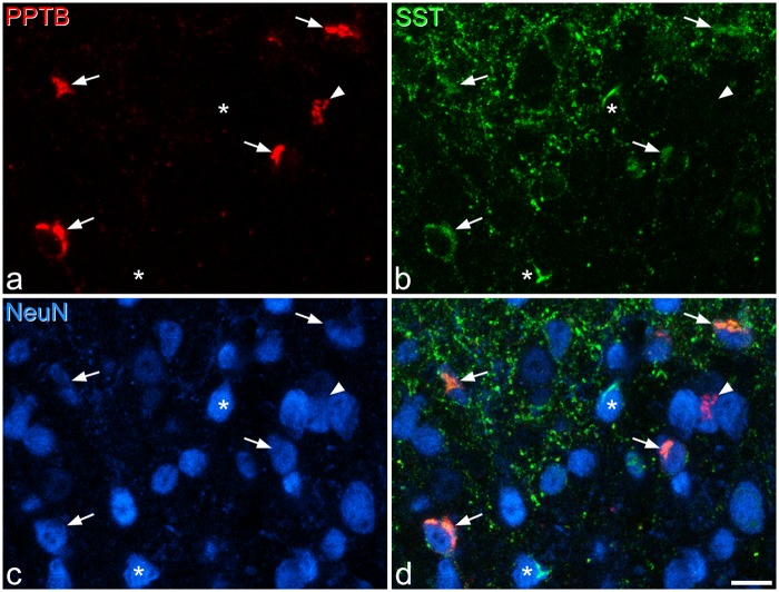
Figure 5.
SST and PPTB immunoreactivity. (a–c) Immunostaining with antibodies against PPTB (red), SST (green), and NeuN (blue) in a maximum intensity projection of three optical sections (1 µm z-spacing) from the lamina II/III border. (d) A merged image. Five PPTB-immunoreactive cells are indicated. Four of these are also SST-immunoreactive (arrows), while the fifth (arrowhead) lacks SST. Two neurons that are SST-immunoreactive but negative for PPTB are marked with asterisks. Scale bar = 10 µm.

