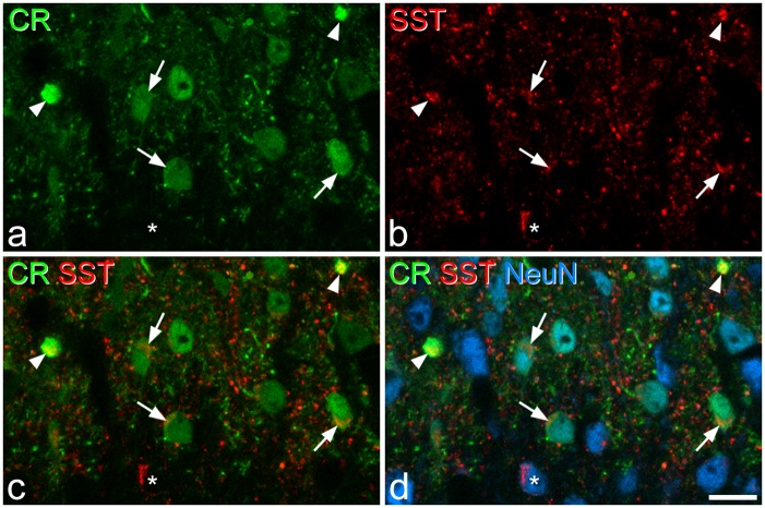
Figure 8.
Calretinin and SST immunoreactivity. (a–c) Immunostaining for calretinin (CR, green) and SST (red) in a single optical section through lamina II. (d) A merged image, which also shows staining for NeuN (blue). Several calretinin-immunoreactive cell bodies are present in the section, and three of these are marked with arrows. Each of these cells show SST-immunoreactivity. The two calretinin-labelled profiles indicated with arrowheads are proximal dendrites of lamina II neurons, and both of these are also SST-immunoreactive. The asterisk marks a SST-immunoreactive neuron that lacks calretinin. Scale bar = 10 µm.

