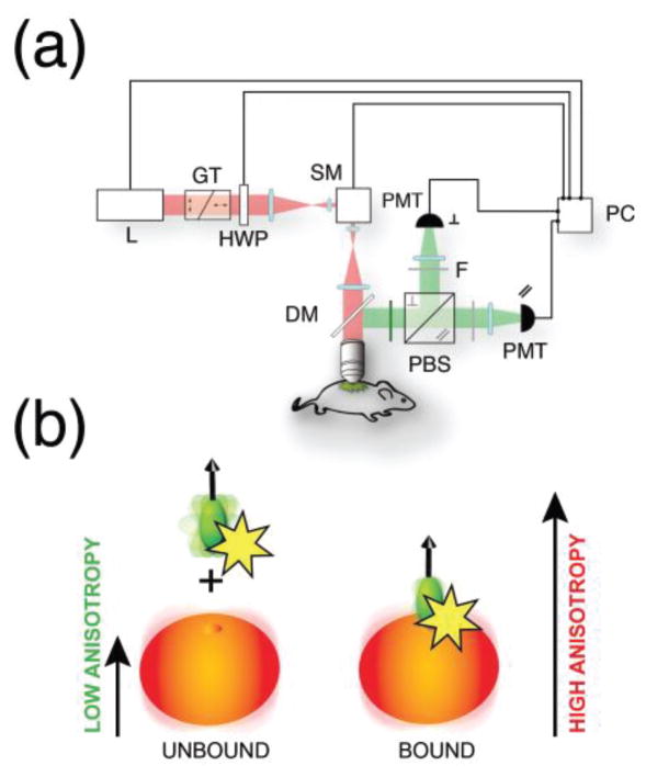
Fig. 3.
(a) Schematic representation of the two-photon fluorescence anisotropy microscopy system. L laser, GT Glan-Thompson polarizer, HWP half waveplate, SM scanning mirror, PMT photomultiplier tube, DM dichroic mirror, PBS polarization beamsplitter, F polarizing filters. (b) Upon binding to their target, fluorescently labeled small molecule drugs present an increase in fluorescence anisotropy due to the larger molecular weigth of the target. Adapted and reprinted with permission from [20].

