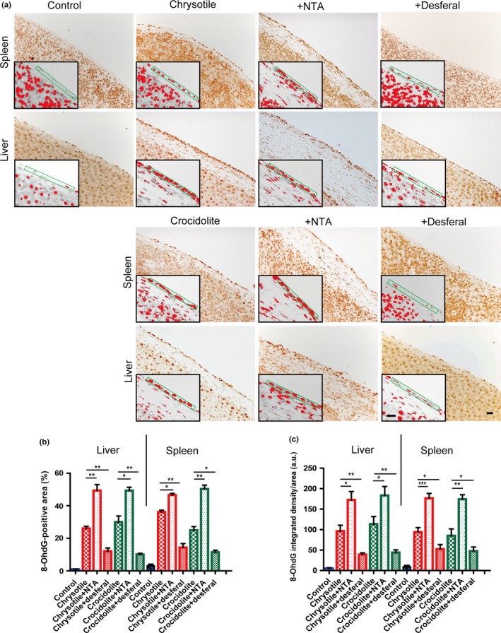
Figure 4.

Immunohistochemical analysis of 8‐hydroxy‐2′‐deoxyguanosine (8‐OHdG) in rat peritoneal mesothelial cells after asbestos injection in the presence or absence of iron chelators. (a) Nuclear immunostaining of surface mesothelial cells were observed 5 weeks after asbestos injection, which was aggravated by nitrilotriacetate (NTA) and ameliorated by desferal (bar = 50 μm). (b,c) Quantitative analysis of immunostained area (b) and integrated density per area (c) (means ± SEM). *P < 0.05; **P < 0.01; ***P < 0.001. a.u., arbitrary unit.
