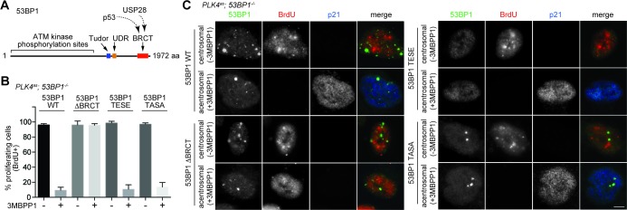
Figure 3. 53BP1 mediates centrosome loss-induced G1 arrest independently to its DNA repair activity.
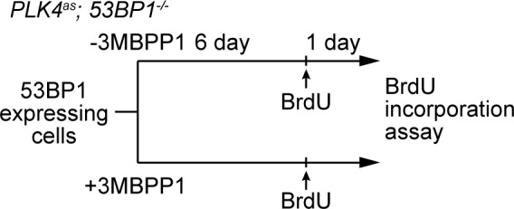
(A) Domain organization of 53BP1. BRCT (BRCA1 carboxy-terminal), UDR (ubiquitylation-dependent recruitment). p53 and USP28 interact with 53BP1 through the tandem BRCT domain. (B) 53BP1ΔBRCT mutant does not rescue the G1 arrest in PLK4as; 53BP1-/- cells after centrosome removal. Wild-type or indicated mutant 53BP1 were mildly expressed under the tetracycline inducible promoter in stable, clonal, centrosomal PLK4as; 53BP1-/- cells (see Materials and methods), during which centrosome loss was induced by 3MBPP1 addition. BrdU was added on day six after 3MBPP1 addition and cells were harvested 24 hr later for BrdU incorporation assay (3MBPP1 treatment for seven days in total). Data are means ± SD. n>150, N = 3. (C) Representative immunofluorescence images of cells in (B) stained with the indicated antibodies seven days after 3MBPP1 treatment. 53BP1 was stained with anti-GFP FITC conjugated antibody. Scale bar, 5 μm.
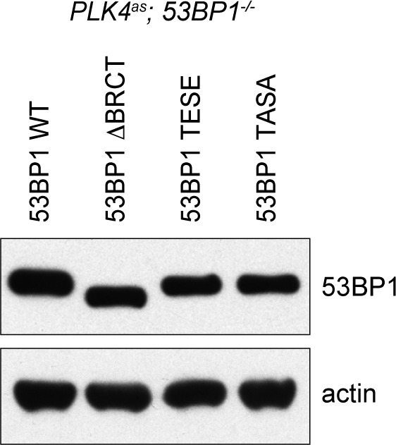
Figure 3—figure supplement 1. Wild type and mutant 53BP1 are exogenously expressed to similar levels in PLK4as; 53BP1-/- cells.

Figure 3—figure supplement 2. Schematic outlining the timeline of drug treatments used.

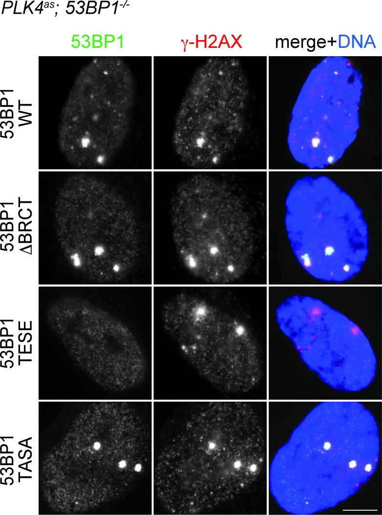
Figure 3—figure supplement 3. DDR function is intact in 53BP1WT, 53BP1ΔBRCT and 53BP1TASA, but not in 53BP1TESE.


