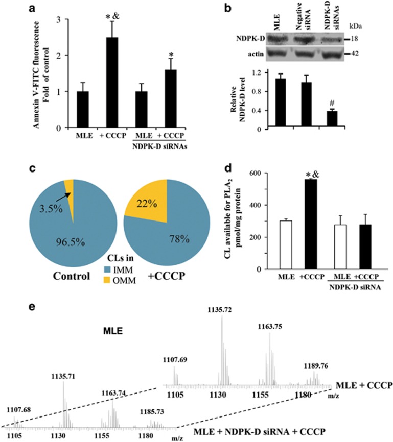
Figure 2.
CCCP induces externalization of cardiolipin in MLE cells. (a) Evaluation of CL in the outer leaflet of OMM using Annexin V-binding assay in w/t and NDPK-D RNAi MLE cells. Cells were treated with 20 μM CCCP for 1 h. Isolated mitochondria were incubated with FITC-labeled Annexin V to stain surface-exposed CL (anionic phospholipids) and then subjected to flow cytometric analysis (FACSCanto, Becton-Dickinson). (b) Knockdown of NDPK-D using siRNA interference in MLE cells. As controls, cells were mock transfected or transfected with non-targeted negative siRNAs. Expression of NDPK-D was evaluated by western blotting. Lower panel shows the relative NDPK-D expression calculated based on densitometry (n=3). (c) LC-MS analysis-based relative contents of CL in OMM and IMM in control and CCCP-treated MLE cells are presented. (d) Evaluation of CL in the outer leaflet of OMM in w/t and NDPK-D knocked down MLE cells using PLA2 treatment with subsequent assay of mono-lyso-CL by LC-MS analysis. (e) Representative MS spectra of mono-lyso CL from MLE cells with normal expression of endogenous NDPK-D or after treatment with NDPK-D siRNAs. *P<0.05 versus control cells without CCCP treatment. #P<0.05 versus cells transfected with non-targeting negative siRNAs. &P<0.05 versus MLE cells transfected with NDPK-D siRNAs under the same condition (20 μM CCCP/1 h)

