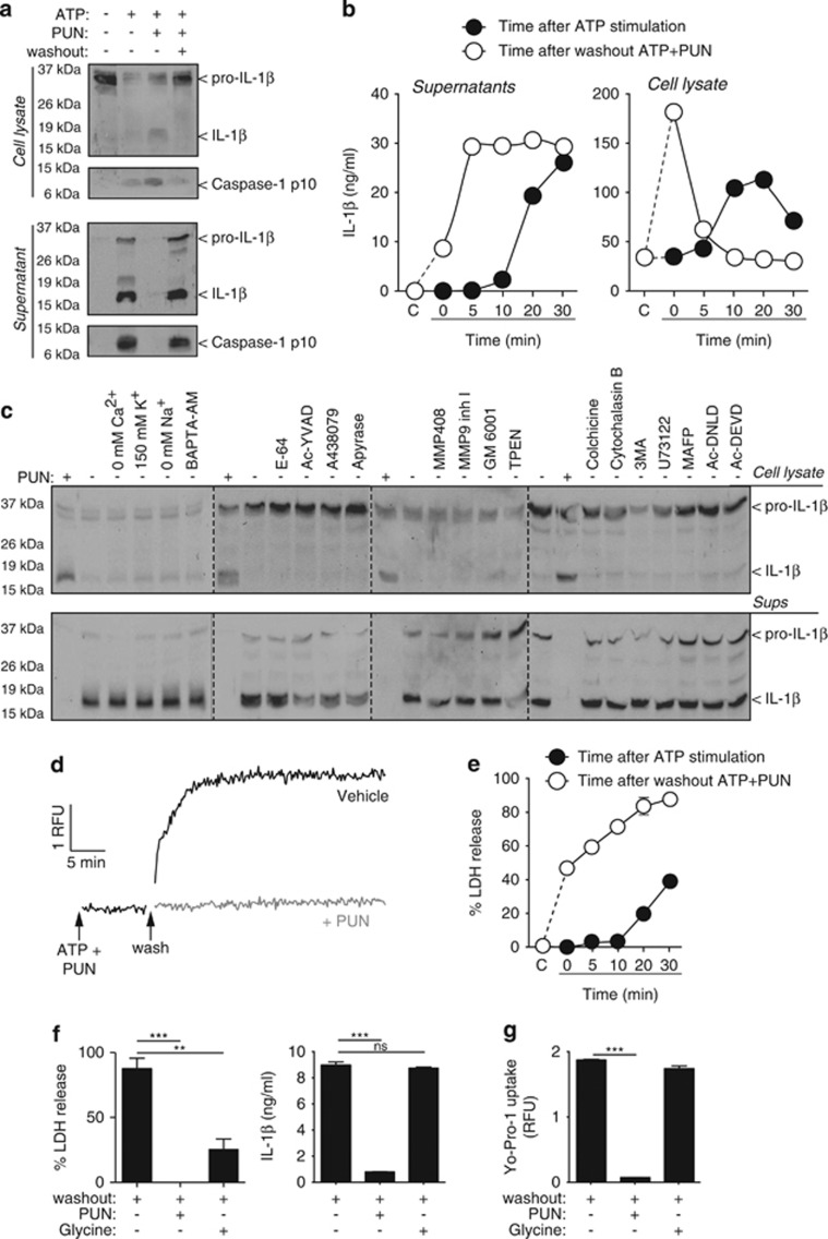
Figure 6.
IL-1β release pharmacology. (a) Immunoblot analysis of cell lysate and supernatant of mouse BMDMs primed with LPS (1 μg/ml, 4 h), followed by no stimulation (−) or stimulation (+) with ATP (5 mM, 20 min) in the absence (−) or presence (+) of punicalagin (PUN; 25 μM) and then washout (+) or not (−) for 20 min. (b) ELISA of IL-1β of cell lysate and supernatant from BMDMs primed as in (a). Measures are taken every 5 min during 30 min of ATP stimulation (5 mM) after priming (black circles) or washout after 30 min stimulation with ATP (5 mM) with punicalagin (PUN; 25 μM) (white circles). (c) Immunoblot analysis of cell lysate and supernatant of BMDMs treated as in (a) and during washout after ATP+PUN cells were incubated with punicalagin (PUN; 25 μM), in a buffer without Ca2+, high K+ (150 mM), with NMDG+ (0 mM Na+), or normal ion buffer with BAPTA-AM (100 μM), E-64 cathepsin inhibitor (50 μM), Ac-YVAD caspase 1 inhibitor (100 μM), A438079 P2X7 antagonist (25 μM), apyrase (3 U/ml), MMP408 (1 μM), MMP9 (0.5 μM) and GM6001 (0.5 μM) metalloprotease inhibitors, TPEN Zn2+ chelator (50 μM), colchicine (50 μM) and cytochalasin B (2.5 μg/ml), 3-MA autophagy inhibitor (6 mM), U73122 phospholipase C inhibitor (10 μM), MAFP phospholipase A inhibitor (10 μM), Ac-DNLD, or Ac-DEVD caspase-3 inhibitors (100 μM). (d and e) Kinetic of Yo-Pro uptake (d) and percentage of extracellular LDH (e) from macrophages treated as in (b). (f) Percentage of extracellular LDH release and ELISA of IL-1β in supernatant from BMDMs treated as in (a) and during washout after ATP+PUN cells were incubated with punicalagin (PUN; 25 μM) or glycine (5 mM). (g) Yo-Pro uptake in BMDMs treated as in (f). **P<0.01; ***P<0.001; n.s., not significant (P>0.05) difference (ANOVA with Bonferroni multiple comparison test)

