. 2016 May 26;5:e12765. doi: 10.7554/eLife.12765

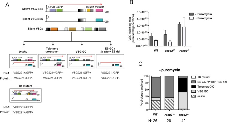
Figure 4. Mutation of TbRECQ2 leads to elevated VSG switching and increased recombination.

(A) Strategy for determining VSG switching mechanisms; adapted from Povelones et al. (2012). The active VSG BES of GFP221hygTK cells is shown, within which the PUR, eGFP, HYG-TK and VSG221 genes are represented as coloured boxes. In addition, one of the ~14 silent BES containing a distinct VSG (turquoise box) is shown, as are multiple silent VSGs elsewhere in the genome (various colours; for convenience these are shown as a single array, but could also be at the telomere of silent mini-chromosomes). 70 bp repeats upstream of the VSGs are denoted by hatched boxes. Different switching strategies allow survival after ganciclovir treatment and can be distinguished by analysis of VSG221 and GFP presence by PCR, and expression of the proteins by western blot (profiles detailed under each mechanism). Switchers that arise by in situ switching, telomere crossover (XO) or VSG gene conversion (VSG GC) can be detected unambiguously, while events that occur by BES gene conversion or in situ switching coupled with BES deletion (ES GC/ in situ+ES del) are indistinguishable. Note, only in situ+ES del reaction is shown, and not ES GC (where all sequence of a silent BES is duplicated and replaces the VSG221 BES); in addition, for VSG GC the silent grey array donor VSG gene is shown as being copied, but the reaction could also use a BES VSG gene. Non-switcher TK mutants can also allow ganciclovir survival. (B) The mean switching rate of GFP221hygTK WT and recq2 mutants (+/- and -/-) was inferred from the mean number of survivors from two experiments, each with three replicates, following treatment with ganciclovir and after culture with (+) or without (-) puromycin; error bars represent standard error of the mean. (C) Profiles of WT and recq2 mutants (+/- and -/-) survivors in the non-puromycin experiments, represented as a percentage of total surviving clones analysed from the two datasets; number of clones (N) analysed is indicated.

DOI: http://dx.doi.org/10.7554/eLife.12765.008

Figure 4.

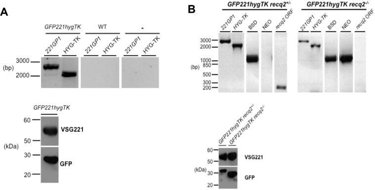
Figure 4—figure supplement 1. Generation of TbRECQ2 mutants in T. brucei GFP221hygTK cells.

Figure 4—figure supplement 1.

(A) Confirmation of the generation of GFP221hygTK cells by PCR and western blot. The upper diagram shows PCR performed on genomic DNA extracted from GFP221hygTK transformants or wild type (WT) cells, testing for integration of the 221GP1 and HYG-TK constructs. Size markers (bp) are shown. The lower diagram shows westerns blots of total protein extract of GFP221hygTK cells probed with rabbit anti-VSG221 antisera or with rabbit anti-GFP antiserum; size markers (kDa) are shown. (B) PCR and western blot analysis of GFP221hygTK recq2 mutants. The upper diagrams show PCR performed on genomic DNA extracted from GFP221hygTK recq2+/- and GFP221hygTK recq2-/- cells, testing for the RECQ2 ORF, integration of the BSD or NEO resistance cassettes, and retention of the 221GP1 and HYG-TK constructs; size markers are shown (bp). The lower diagram shows westerns blots of total protein extract of GFP221hygTK recq2+/- and GFP221hygTK recq2-/- cells cells probed with rabbit anti-VSG221 antisera or with rabbit anti-GFP antiserum; size markers (kDa) are shown. In all gels, gaps indicate that lanes have been aligned in this figure after excision from multiple gels/membranes, or from disparate parts of the same gel/membrane.
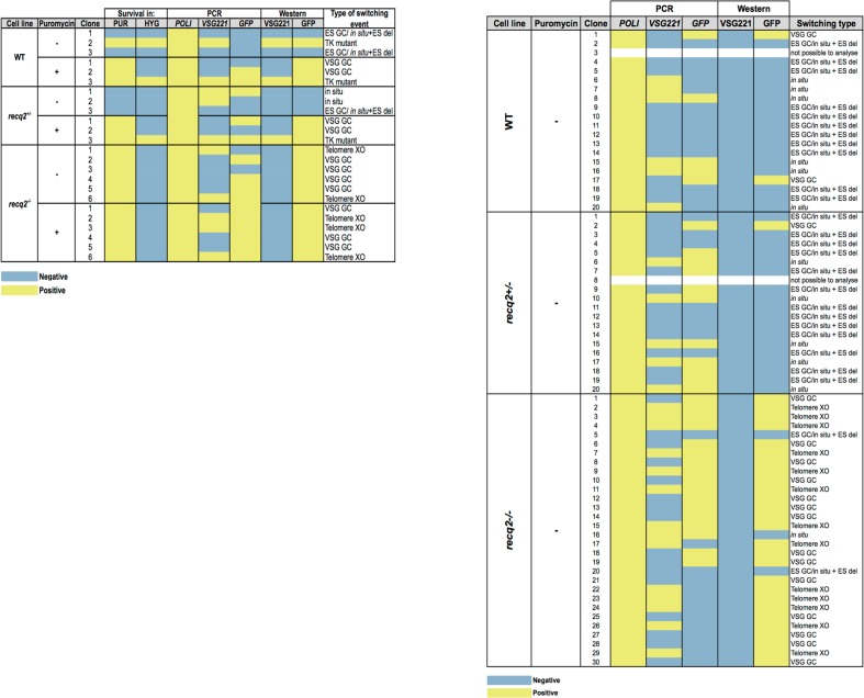
Figure 4—figure supplement 2. Summary table of ganciclovir survival mechanisms.

Figure 4—figure supplement 2.

Summary of PCR and western analysis of clones from the first (left) and second (right) ganciclovir survival experiments. GFP221hygTK wildtype (WT), GFP221hygTK recq2+/- and GFP221hygTK recq2-/- cells are shown, as are whether or not clonal survival in ganciclovir was conducted in the presence (+) or absence (-) of puromycin. ‘Survival in’ denotes whether or not the clones that were recovered could grow in the presence of puromycin (PUR; 1 μg.mL-1) or hygromycin (HYG; 10 μg.mL-1). The following columns summarise the results of PCR to assay for the presence or absence of RNA POLI, GFP and VSG221 genes, and westerns to detect expression of GFP or VSG221; blue indicates a negative result and yellow indicates a positive result, while completely white rows indicate clones that could not be analysed, either due to absence of detectable genomic DNA or of protein in the cell lysate. The final column summarises the inferred strategy for survival, as detailed in Figure 5.