
FIGURE 2.
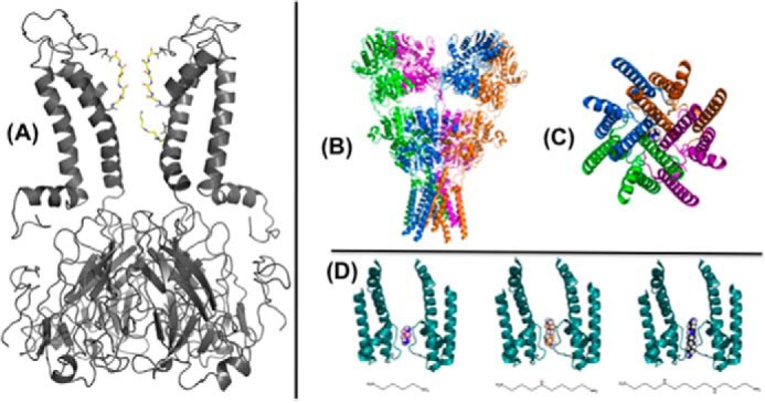
Polyamine interaction with ion channels. A, spermine is depicted in a deep binding site in the inner cavity of a molecular model of the Kir6.2[N160D] mutant channel, generated as a homology model based on an open conformation model of KirBac1.1 (98) with this blocker configuration generated using AutoDock as described previously (54), and selected based on functional studies of Kir6.2[N160D] and Kir2.1 channel (55). B, side view of the crystal structure of the full-length ionotropic glutamate receptor, GluA2 (Protein Data Bank (PDB) ID 3KG2), with a single spermine molecule manually positioned in the transmembrane pore region. C, view of the GluA2 transmembrane domain from the intracellular side showing a single spermine in the pore region. D, two subunits (A/C) of the Bacillus cereus NaK channel pore (PDB ID 3E86) with putrescine (left), spermidine (middle), and spermine (right) docked in the selectivity filter (66).