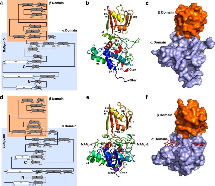
FIGURE 2.
Overall structure of BaSpoIID (top) and CdSpoIID (bottom). a and d, topology diagrams of BaSpoIID and CdSpoIID, respectively, with the highlighted α-helix rich α-domain in the slate box and the β-strand rich β-domain in the orange box. The three α-helices comprising the core of the α-domain are shown in white. b and e, ribbon diagrams of BaSpoIID and CdSpoIID, respectively, rainbow-colored from blue at the N terminus to red at the C terminus. e, two NAG3 molecules are represented as green sticks. c and f, surface representations of BaSpoIID and CdSpoIID, respectively, with the characteristic fist-like shape, with the α-helix rich α-domain (the hand) colored in light blue and the β-strand rich β-domain (the arm) in orange. f, the two NAG3 molecules are represented as red sticks in the main groove of the α-domain.

