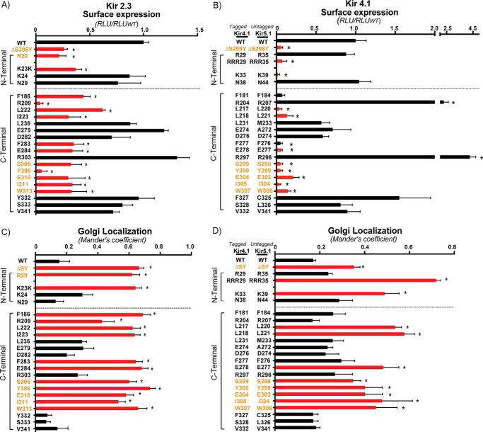
FIGURE 4.
Golgi export sequence determinants of Kir2.3 and Kir4.1 elucidated by structure-guided mutagenesis. Residue candidates for the Golgi export sequence determinants were subjected to alanine replacement mutagenesis. A and B, cell surface expression of external HA-tagged Kir2.3 and Kir4.1 channels as quantified by surface HA antibody binding (n = 3). RLU, relative light unit. C and D, quantification of Kir2.3 and Kir4.1 co-localization with the Golgi marker GM130 (n = 30 cells from three individual transfections, the fraction of co-localized channel is presented as Mander's coefficient). Red bars, Golgi export sequence determinants; orange text, previously identified residues in the Kir 2.1 Golgi export signal. *, p < 0.05 by one-way randomized ANOVA followed by Dunnett's post hoc test.

