
FIGURE 4.
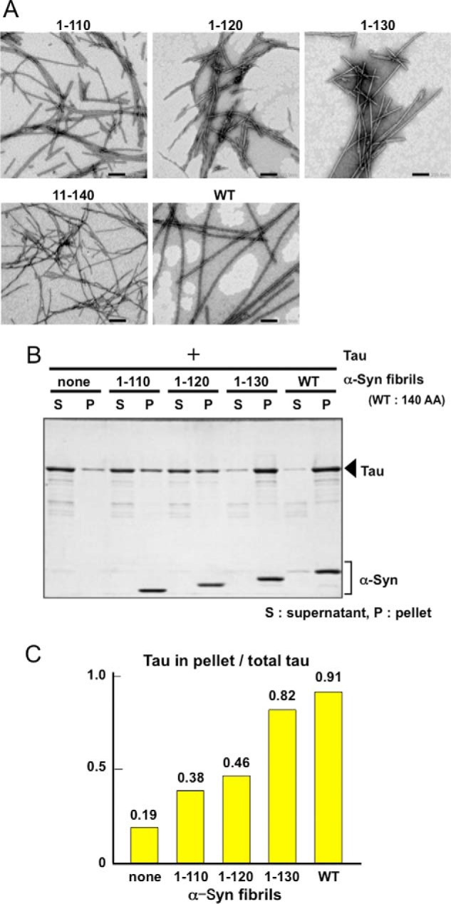
Interaction of Tau with full-length or C-terminally truncated α-synuclein fibrils. A, negative staining of electron microscopy of α-synuclein fibrils consisting of C-terminally truncated (1–110, 1–120, and 1–130), N-terminally truncated (11–140), and full-length wild-type α-synuclein (WT). B, α-synuclein (α-Syn) fibrils consisting of full-length wild-type or C-terminally truncated α-synuclein were incubated with Tau, and the binding was analyzed by means of spin-down assay. α-Synuclein fibrils of each type (22.1 μm) were mixed with an equal volume of 7.4 μm Tau and centrifuged. The fibril-bound Tau recovered in the pellet and the unbound Tau in the supernatant were analyzed by SDS-PAGE and Coomassie Brilliant Blue (CBB) staining. C, relative ratio of bound Tau in pellet to the total Tau is shown.