
FIGURE 6.
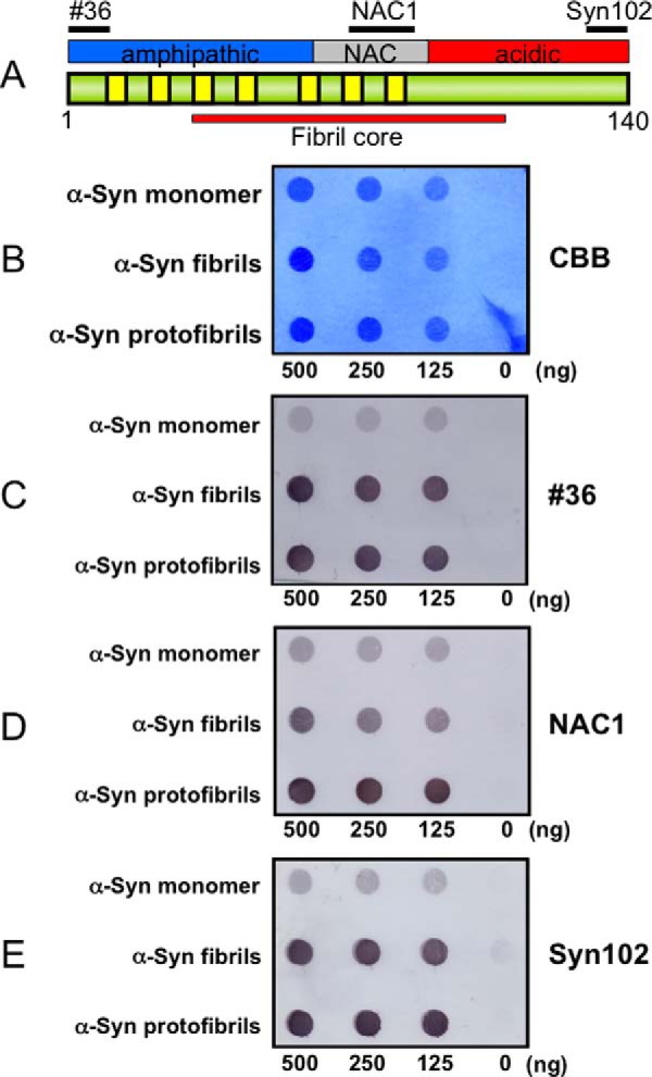
Dot blot assay of α-synuclein monomer, fibrils, and protofibirils with various anti-α-synuclein antibodies. A, schematic diagram of the domain structure of the α-synuclein (α-Syn) molecule. Epitopes of antibodies 36 (residues 1–10), NAC1 (residues 75–91), and Syn102 (residues 131–140) used in the dot blot assay are shown. B, Coomassie Brilliant Blue (CBB) staining of α-synuclein monomer, fibrils, and protofibrils dotted on a nitrocellulose membrane. C, immunodetection with an antibody to N terminus of α-synuclein (#36). D, immunodetection with an antibody to the mid-portion of α-synuclein (NAC1). E, immunodetection with an antibody to C terminus of α-synuclein (Syn102).