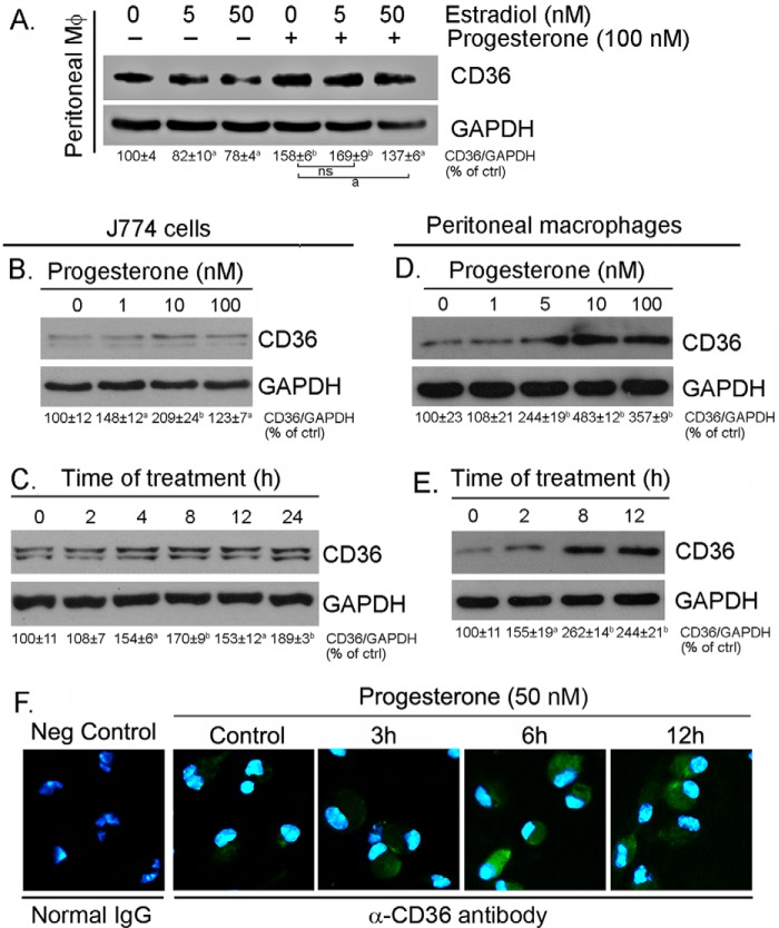
FIGURE 1.
Progesterone induces macrophage CD36 protein expression. A, peritoneal macrophages were isolated from female C57BL/6 mice and treated with estradiol at the indicated concentrations or plus 100 nm progesterone overnight. a, p < 0.05; b, p < 0.01 versus control in the corresponding groups; ns, not significantly different (n = 3); ctrl, control. B–E, J774 cells at ∼95% confluence or peritoneal macrophages isolated from female C57BL/6 mice in serum-free medium were treated with progesterone at the indicated concentrations overnight (B and D) or with 50 nm progesterone for the indicated times (C and E). After treatment, cellular proteins were extracted and used to determine CD36 expression by Western blotting. All bands in the Western blots were scanned, and the density of the target band normalized by GAPDH was calculated with statistical analysis. a, p < 0.05; b, p < 0.01 versus control in the corresponding groups (n = 3). F, peritoneal macrophages isolated from female C57BL/6 mice were treated with 50 nm progesterone for the indicated times, followed by determination of CD36 protein expression in intact cells by immunofluorescent staining. Neg Control, negative control. Cells were added with normal rabbit IgG.

