
FIGURE 3.
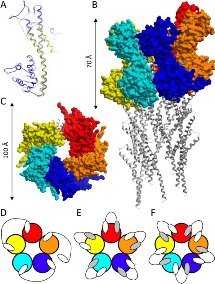
PcrV modeling and Nanobody binding modes. A, homology model of PcrV (blue), superimposed to the C-terminal helices of the S. flexneri needle subunit (yellow). Shown are side (B) and top (C) views (not drawn to the same scale) of the pentameric PcrV model complex (surface representation) on the tip of a T3SS needle (ribbon representation). Shown are theoretical binding modes of bivalent (D) and biparatopic (E and F) Nanobodies to a pentameric PcrV ring (not drawn to the same scale). Individual PcrV protomers are shown in different colors. White and gray ovals, Nanobody building blocks binding to distinct epitopes on a PcrV monomer.