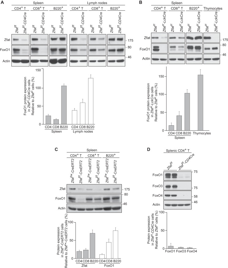
FIGURE 1.
Decrease in FoxO1 protein in Zfat-deficient T cells. A–D, immunoblot analysis of the indicated proteins on peripheral CD4+ T, CD8+ T, and B220+ cells or thymocytes from the indicated genotype mice. Actin was used as a loading control. Levels of protein expression were quantified by densitometry and normalized to actin levels. C, Zfatf/w-CreERT2 and Zfatf/f-CreERT2 mice were treated with tamoxifen for 3 days. A–C, data are representative of two or three independent experiments.

