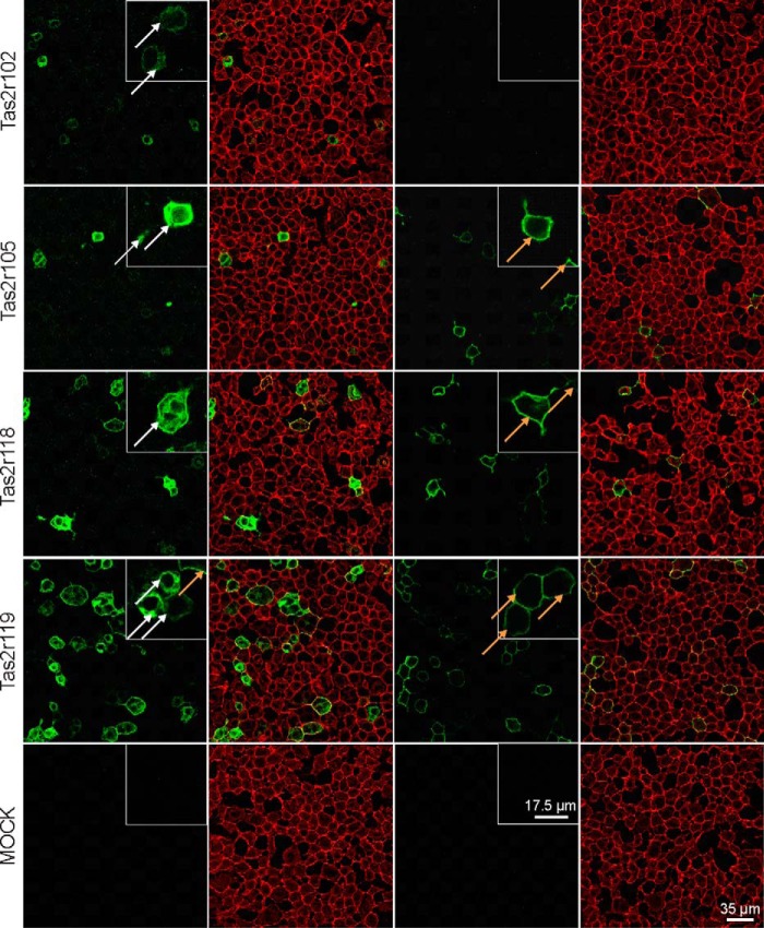
FIGURE 4.
Confocal fluorescence images of HEK293T-Gα16gust44 cells transiently transfected with cDNAs of mouse bitter taste receptors. Cells were transfected with Rho-tagged Tas2r constructs and underwent immunostaining with Rho antibody (green) either after (two left panels) or before fixation (two right panels). The cell surface was visualized by biotin-conjugated concanavalin A and streptavidin-conjugated Alexa Fluor 633 (red). Taste receptors located on the cell surface appear yellow in the overlay and are exemplarily indicated by orange arrows. Receptors without surface expression are exemplarily indicated by white arrows.

