FIGURE 1.
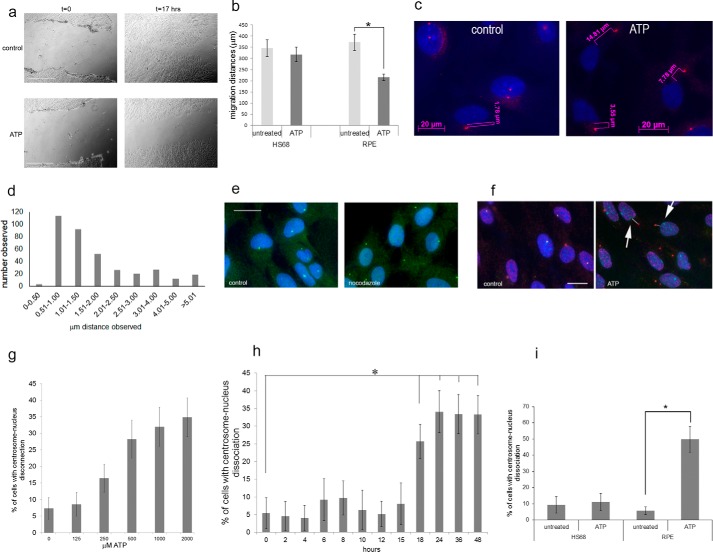
Extracellular ATP inhibits cell migration and causes centrosome-nucleus separation. The effect of ATP on cell migration was studied using the scratch assay. a, examples of phase-contrast images of scratch-damaged monolayer RPE cells immediately and 17 h after the scratch with and without 2 mm ATP treatment. Scale bars = 500 μm. b, quantitation of results from eight scratch assays for each cell line. c, we analyzed centrosome-nucleus separation in untreated RPE cell cultures (control) and in cells treated with ATP (ATP) as follows. Cells were fixed and stained with an antibody to ninein to detect the centrosome (pink) and DAPI to stain the nucleus (blue). Images were obtained with immunofluorescence microscopy. We analyzed centrosome-nucleus distances as shown using Axiovert software. d, the distance between the centrosome and nucleus was measured in RPE cells growing under normal conditions. Data were collected from 360 cells. The distribution of distances was analyzed and is shown. e, nocodazole causes centrosome-nucleus separation. Control, untreated RPE cells; nocodazole, RPE cells treated with 0.5 μm nocodazole for 3 h. Anti-pericentrin was used to show centrosomes, and anti-β-tubulin was used to reveal microtubules. DAPI stains nuclei. Images were obtained with immunofluorescence microscopy. Scale bar = 20 μm. f, RPE cells were analyzed without treatment (control) or after treatment with 2 mm ATP (ATP) for 24 h, fixed, and stained with human autoimmune serum (M4491) and an antibody to ninein, both to visualize the centrosome, and with DAPI to stain nuclei. Images were obtained with immunofluorescence microscopy. Arrows point to examples of cells with distanced centrosomes and nuclei, schematically indicated by the white lines. Distances were measured. Scale bar = 20 μm. g, results of ATP dose-response experiments. RPE cells were treated with the indicated concentrations of ATP for 16 h. Data were collected from three independent assays, for each of which the centrosome-nucleus distance was measured in 300 cells. h, ATP treatment time course. RPE cells were treated with 1 mm ATP for the indicated times, and centrosome-nucleus distances were measured. Data were collected from three independent assays, for each of which at least 300 cells were analyzed. i, HS68 cells and RPE cells were analyzed untreated or after treatment with 1 mm ATP, and distances between the centrosome and nucleus were measured. Data were collected from three independent assays, for each of which at least 300 cells were counted. *, p < 0.05.

