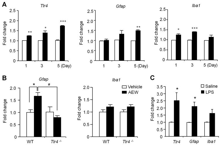
Figure 7. Quantitative RT-PCR shows the expression of Tlr4 and glia-related genes in cervical spinal cord dorsal horn (C3–C4) after AEW treatment on the back skin or intrathecal LPS injection.
(A) Relative expression levels of Tlr4, Gfap, and Iba1 1, 3, and 5 days after AEW treatment. *P<0.05, **P<0.01, **P<0.001, compared with corresponding vehicle control, n = 4 mice per group. (B) Relative expression levels of Gfap and Iba1 in the dorsal horn of WT and Tlr4−/− mice 5 d after AEW treatment. *P<0.05; #P<0.05, n = 5 mice per group. Note that dry skin induces Tlr4-dependent Gfap expression. (C) Relative expression of Tlr4, Gfap, and Iba1 in cervical spinal cord dorsal horn 24 h after intrathecal LPS (10 μg) injection. Data are presented as means ± S.E.M. *P<0.05, compared with corresponding vehicle control, Student’s t test; n = 4 mice per group.

