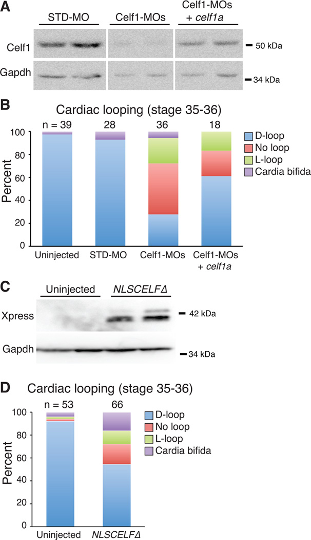
Fig. 6. Morpholino-mediated knockdown of Celf1 can be ameliorated by restoration of Celf1 expression, and mimicked by repression of nuclear Celf activity.
Xenopus laevis embryos were injected either with morpholino oligonucleotides (STD-MO, control morpholino; Celf1-MOs, a mix of two celf1-targeting morpholinos) ± celf1a RNA (A, B), or with RNA encoding a dominant negative CELF protein (NLSCELFΔ; C, D) at the 2-4-cell stage and compared to uninjected controls at stage 35–36. (A) Celf1 protein levels were determined by western blotting. Two embryos per group are shown. Lanes shown are from the same blot. (B) Cardiac looping and the incidence of cardia bifida following knockdown and restoration of Celf1 were evaluated by whole-mount immunohistochemistry using antibodies against meromyosin (MF20) or Tnnt2 (CT3). (C) Dominant negative protein expression was confirmed at stage 26 by western blotting for its Xpress epitope tag. Two embryos per group are shown. (D) Cardiac looping and fusion defects were evaluated in embryos expressing the dominant negative CELF protein by whole mount immunohistochemistry against meromyosin. D-loop, dextro-loop; L-loop, levo-loop.

