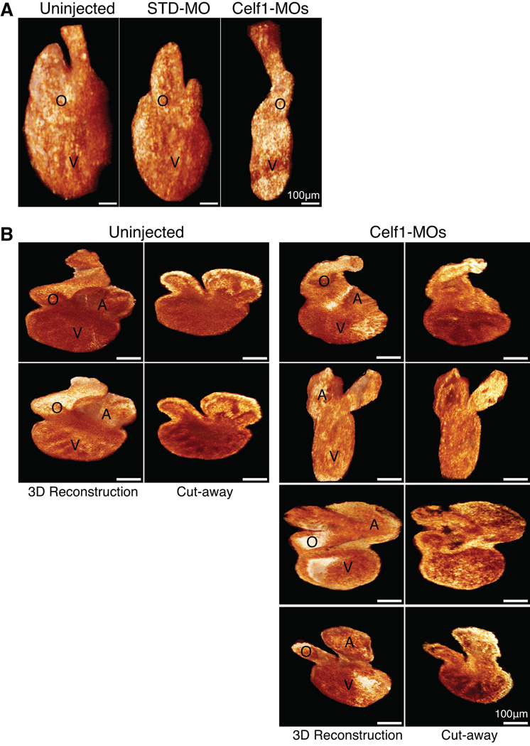
Fig. 8. Cardiac dysmorphia following morpholino-mediated knockdown of Celf1 in Xenopus laevis was imaged in situ and ex vivo at stage 46.
The hearts of uninjected (n = 9), STD-MO-injected (n = 7), and Celf1-MOs-injected (n = 8) embryos at stage 46 were imaged in situ by optical coherence tomography (OCT), and then removed and imaged by optical coherence microscopy (OCM). (A) Representative examples of three-dimensional reconstructions of OCT sections through the hearts are shown. General dysmorphia of the heart and outflow tract can be seen following Celf1 depletion; the atria cannot be resolved in these reconstructions. (B) Representative examples of hearts imaged ex vivo using OCM, in intact three-dimensional reconstructions (left) and in cut-away views (right). Multiple hearts from Celf1-MOs-injected embryos are presented to demonstrate the range of dysmorphia observed. Heart reconstructions are shown at equivalent orientations for the purpose of comparison; this orientation does not necessarily match the orientation of the hearts in situ. A, atrium; O, outflow tract; V, ventricle.

