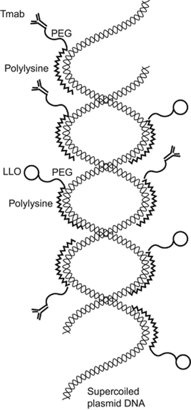
Figure 9.
Illustration of a single Tmab-targeted DNA nanocomplex. A supercoiled double-helical plasmid DNA molecule is shown in the interior, decorated with numerous Tmab-PEG-PL molecules and LLO-PEG-PL molecules on the exterior. The PEG found in each of these attachments is envisioned as a flexible arm protruding out from the PL bound to the supercoiled DNA molecule. Small-angle neutron scattering studies of mono-PEGylated conjugates, in which PEG is covalently linked to a protein, assume a dumbbell configuration rather than a shroud configuration.23 Presumably this would leave the Tmab positioned in such a way as to be able to bind to the Her2 receptors in the plasma membranes of targeted cells. The molecular weights of the individual components in the nanocomplex are as follows: plasmid DNA=4.7 kb (pEGFP-N3) or 5.9 kb (NanoLuc); Tmab=145.5 kDa; LLO=58 kDa; PL=37 kDa; and PEG=5 kDa. The overall size of the nanocomplex is 150–250 nm. Her2, human epidermal growth factor receptor 2; LLO, Listeriolysin O; PEG, polyethylene glycol; PL, polylysine; Tmab, trastuzumab.

