
Figure 2.
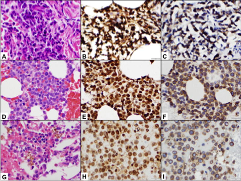
Patient #222-023. Prior to the therapy, the patient had multiple skin infiltrates. Skin biopsy detected dense perivascular infiltrate composed of intermediate to large neoplastic cells with irregular nuclear contours, slightly clumped chromatin, indistinct nucleoli, and scant to moderate pink cytoplasm (A, H&E, ×500). The neoplastic cells demonstrated strong and uniform HIF-1α expression (B, IHC, ×500) and strong CAIX expression (C, IHC, ×500). Bone marrow biopsy prior to therapy demonstrated hypercellular (70–80%) bone marrow with sheets of immature cells (D, H&E, ×500). Immunohistochemical studies detected strong and uniform HIF-1α expression (E, IHC, ×500); CAIX expression was somewhat weaker and seen in about 60–70% of cells (F, IHC, ×500). After 1 cycle of evofosfamide at 550mg/m2 (C1D40), patient had complete resolution of leukemia cutis and reduction of BM blasts from 69 to 21% (G, H&E, ×500) with corresponding decrease in fraction of BM HIF-1α-expressing cells (95% vs 60%, H, IHC, ×500) and no change in CAIX expression (70% vs 80%, I, IHC, ×500).