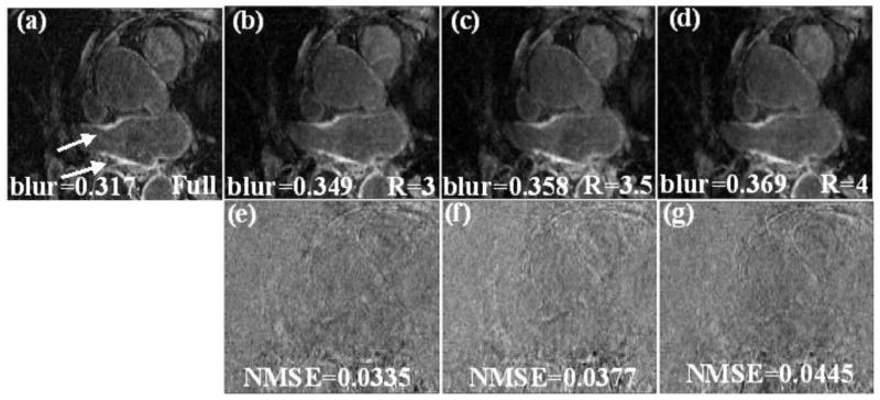
Fig 2.
(a) Cropped LA region in one slice from a fully sampled image, the arrows point to the enhancement in the LA wall. (b), (c) and (d) Reconstruction using the proposed method with “adding noise back” for R=3, R=3.5 and R=4 respectively (using eq. (6) and (3)). The blur metric for the truth and the reconstructed images are reported along with the images. (e), (f) and (g) Difference image between the truth and the images reconstructed in (b), (c) and (d) respectively. The MSE of the individual slice is shown along with the difference image.

