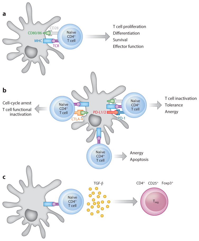
Figure 1.
Tolerance by antigen (Ag) presentation without costimulation. Antigens are internalized by antigen-presenting cells (APCs), and after processing, a peptide of 7–10 amino acids or 14–20 amino acids can be presented on major histocompatibility complex (MHC) I or II, respectively, for recognition by the T cell receptor (TCR) on T cell subsets. The plasticity of T cell subsets enables their response resulting in either immunity or the induction of tolerance. (a) Immunity. The T cell clones mediating the undesired response contain a TCR that recognizes the Ag presented by an APC presenting the MHC/Ag complex (signal 1). The interaction between costimulatory factors present on APCs and cognate receptors on T cells is also needed to cause T cell activation (signal 2). The final stage of T cell activation involves the interaction of soluble mediators (such as interleukins or other factors) with receptors on the T cell surface (signal 3). (b) Blockade of the positive costimulatory CD28/B7 signaling axis between T cells and APCs by the cytotoxic T lymphocyte antigen 4 (CTLA-4)–Ig fusion protein serves as a constitutive regulatory control switch, and it has been exploited as a tolerogenic regimen for autoimmunity treatment. Programmed death 1 (PD-1) signaling on T cells by its ligands PD-L1 and PD-L2 represents another regulatory mechanism of immune responses, and it has been extensively studied in the context of autoimmunity and T cell exhaustion and deletion. Selective blockade of the PD-1/PD-L1 signaling axis can disrupt tolerance induction by blocking T cell receptor stop signals, thereby decreasing T cell motility and increasing physical interactions with APCs, resulting in enhanced immune responses. (c) Induction of regulatory T cells (Tregs) as a mechanism to achieve Ag-specific immunoregulation. In the presence of transforming growth factor β (TGF-β) and interleukin-2 (IL-2), naïve T cells can be differentiated into Tregs that release immunosuppressive cytokines such as IL-10 to diminish the induction and proliferation of effector T cells.

