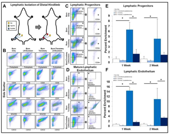
Figure 2. Lymphatic isolation of the distal hindlimb results in abrogation of lymphagiogenic markers at the site of tenotomy.
A. Model of lymphatic isolation of the distal hindlimb via combined popliteal and inguinal lymph node excision. B. Lymphatic isolation reduces the enrichment of lymphatic endothelial and progenitor markers at the site of tenotomy. C. Flow panel identifying lymphatic progenitor cells (LPCs: CD34+/VEGFR3+/CD133+). D. Flow panel identifying mature lymphatic endothelium (MLE: CD31/+Podoplanin+/VEGFR3+/CD133−). E. Quantitative analysis of lymphatic progenitors in the distal hindlimb in the presence or absence of injury models. F. Quantitative analysis of lymphatic endothelium in the distal hindlimb in the presence or absence of injury models. All error bars represent standard deviation. Black asterisks on graph represent p<0.05.

