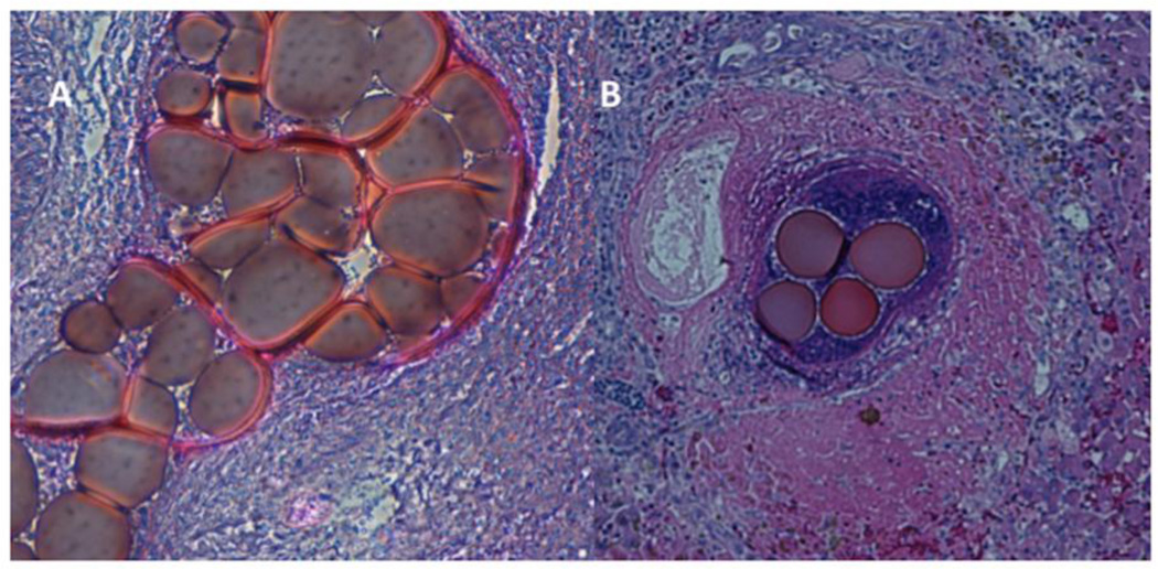
Figure 1.
Pathology images of H&E-stained slides of tumours treated with 70–150 µm DEB.
A. Cluster of 70–150µm DEB inside a tumour-feeding arteriole, which they almost completely obliterate, at the tumour centre (magnification × 40).
B. Cluster of 70–150µm DEB inside a tumour-feeding arteriole, at the tumour periphery (magnification × 25), in a different animal than in Figure 1A. Tumour is identified in the left section of this image, with characteristic tumour cell necrosis (purple). Healthy hepatic parenchyma is identified at the right of this image. There is also inflammatory reaction (purple) at the area surrounding the DEB.

