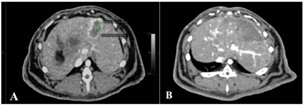
Figure 5.
Axial contrast-enhanced CT images of rabbit liver, demonstrating VX2 liver tumour response to intra-arterial injection of 70–150 µm DEB.
A. Baseline axial contrast-enhanced (late arterial phase) CT images of rabbit liver, demonstrating a VX2 tumour (green calipers), measuring 2.13 × 1.22 cm, at 14 days following liver VX2 tumour implantation and 24 hours before treatment with intra-arterial injection of 70–150 µm DEB. Note the hypovascular necrotic core, already present at 14 days.
B. Axial contrast--enhanced (late arterial phase) CT images of rabbit liver, 7 days after treatment with intra-arterial injection of 70–150 µm DEB. Note there is no visibly enhancing tumour, rather a hypovascular wedge-shaped area corresponding to the area where the VX2 tumour is located. This response was deemed complete response, according to the modified RECIST.

