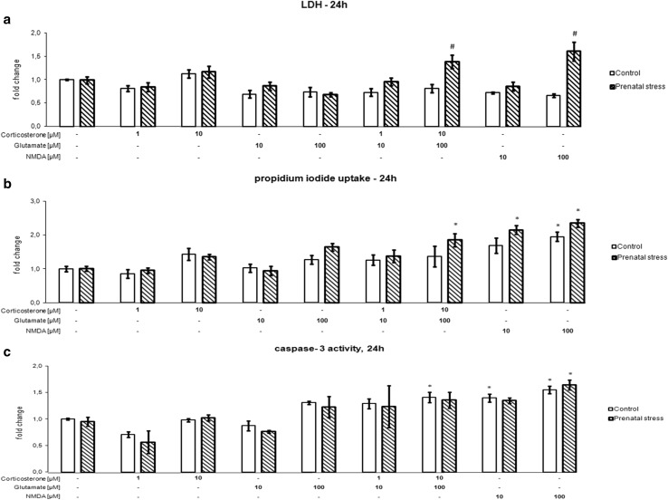
Fig. 1.
The effect of 24 h of exposure of hippocampal organotypic cultures to corticosterone, glutamate, corticosterone with glutamate, and NMDA on LDH release (a), propidium iodide uptake (b), and caspase-3 activity (c). The results are shown as a fold change relative to control cultures exposed to the appropriate vehicle and are expressed as the mean ± SEM. The significance of differences between the means was evaluated by Tukey’s post hoc tests following a factorial analysis of variance (ANOVA). *p < 0.05 versus control cultures; # p < 0.05 versus equally treated cultures from prenatally stressed rats; n = 8

