Fig. 3.
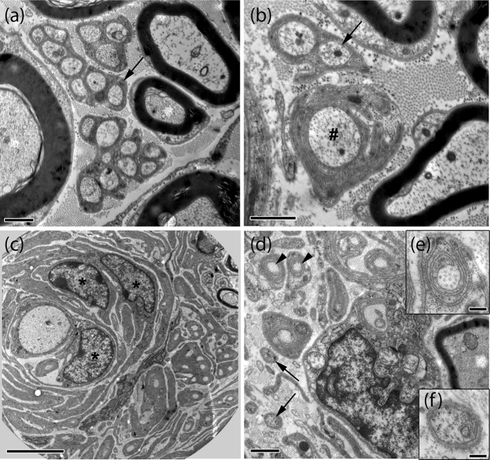
Electron microscopy reveals multiple signs of degeneration in crushed and uncrushed nerves of P0-Cre;Nefh-Cre;Nf2fl/+ mice. Representative electron microscopic images of intact (a, b) and crushed (c–f) sciatic nerves (8 months post-injury) taken from P0-Cre;Nefh-Cre;Nf2fl/+ mice. While myelinated axons and Remak bundles of various size (arrows in a, b) are frequent in intact nerves, myelinated axons and Remak bundles appear rarely in crushed nerves (c, d). Non-myelinated axons were mostly seen as single axons (arrows in d, f). Signs of degeneration such as collagen pockets (arrowhead in d, e), abnormally thin myelin sheath and layers of Schwann cell processes enwrapping axons (c), bands of Bungner and axons with irregular myelin sheaths (hash in b) were mostly present in crushed, but, to a minor extent, also in intact nerves. In addition, accumulations of Schwann cells could be observed (asterisks in c). Scale bars in a, b, d represent 1 μm. Scale bar in c 5 μm. Scale bars in e, f represent 0.2 μm

