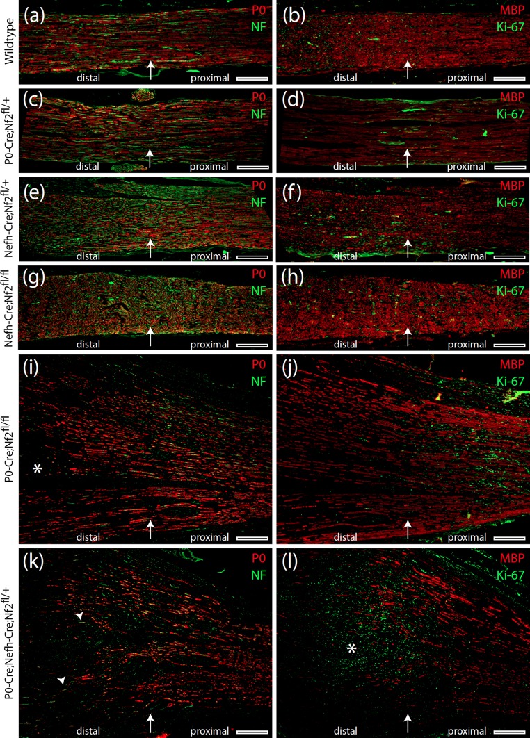
Fig. 4.
Severe re-myelination defect in P0-Cre;Nefh-Cre;Nf2fl/+ mice following nerve crush. a–l Immunohistochemical stainings of longitudinal sciatic nerve sections prepared from indicated genotypes 8 months after crush injury. Immunolabeling of P0, neurofilaments, MBP and Ki-67 indicates Schwann cell differentiation, axonal fibres, myelination and cell proliferation, respectively. Arrow in each image shows the position of the nerve crush. Orientation of nerves is stated as ‘distal’ and ‘proximal’. Asterisk in i emphasizes an area of defective re-myelination. Arrowheads in k indicate axons devoid of any myelin sheath. Asterisk in l marks concentration of Ki-67-positive cells at the edge of intact myelination. Scale bars represent 200 μm

