Fig. 5.
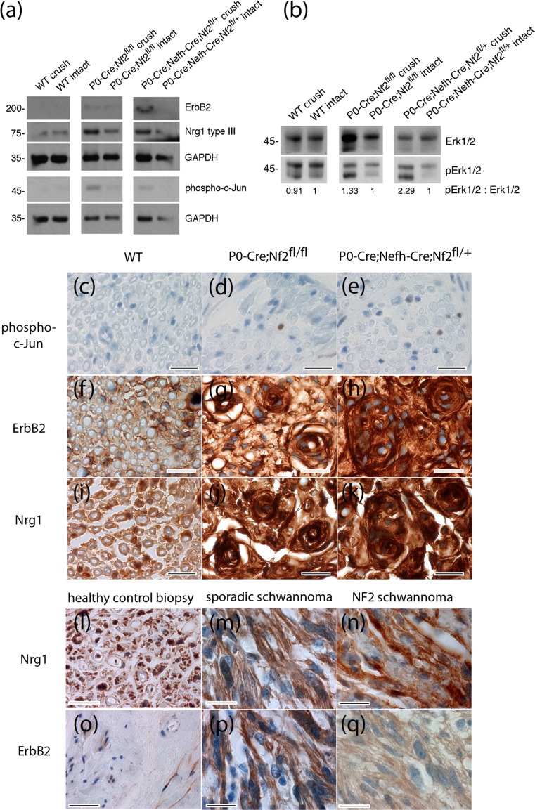
Signaling and protein expression changes in sciatic nerve lysates. a, b Immunoblot of sciatic nerve lysates (pooled tissue from at least three different animals per indicated genotype was prepared from crushed and intact sciatic nerves 8 months after crush injury). a Immunoblot for receptor tyrosine kinase ErbB2, Neuregulin 1 type III (Nrg1 type III), phospho-c-Jun and GAPDH as loading control (n = 3). For full-length blot see Supplementary Fig. 9. b Immunoblot for phospho-Erk1/2 (pErk1/2). Total protein amount of Erk1/2 served as loading control. Densitometric quantification of pErk1/2: Erk1/2 ratio was normalized to intact nerve tissue for each genotype (n = 3). The observed increase in total Erk after crush injury in some genotypes (see also Supplementary Fig. 10) remains unexplained but does not effect the ratio between phospho-Erk and overall Erk. For full-length blot see Supplementary Fig. 10. c–k Sciatic nerve cross sections of indicated genotypes 8 months after crush injury were immunolabeled (brown color) for phospho-c-Jun, as a marker of cellular de-differentiation (c–e), ErbB2 (f–h) and Neuregulin 1 (i–k). Cell nuclei are visualized in blue. Scale bars represent 20 μm. l–q Human tissue sections taken from healthy sural nerve biopsies, as well as sporadic and NF2-associated schwannomas, were immunolabeled (brown color) for Neuregulin 1 (l–n) and ErbB2 (o–q). Cell nuclei are visualized in blue. Scale bars represent 20 μm

