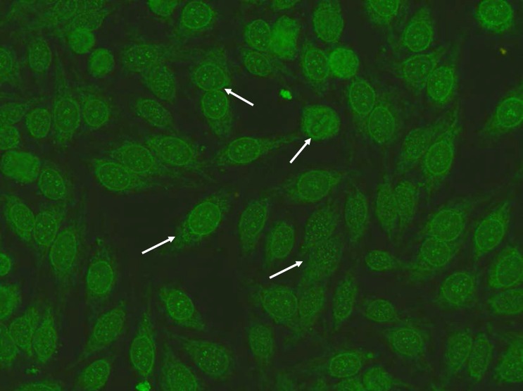
Figure 2.
Patient II IIF Hep-2 staining pattern: Anti-centromere A staining pattern: Rather uniform discrete speckles located throughout the entire nucleus. Telophase and metaphase cells always show these speckles in the condensed chromosomal material. Punctate nuclear membranous pattern: focusing through the nucleus can be seen on the surface of the entire nucleus. A similar pattern is seen in telophase and in metaphase the fluorescence is diffusely localized throughout the cytoplasm

