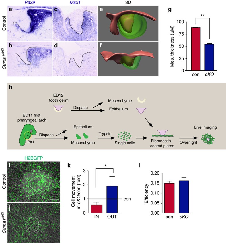
Figure 5. αE-catenin is required for the epithelium-induced mesenchymal condensation during incisor development.
(a–d) In situ hybridization for Pax9 and Msx1 in control and Ctnna1cKO. In control embryos, Pax9 and Msx1 are expressed in the mesenchyme of the tooth germ at E13.5. In Ctnna1cKO, the expression of these genes is reduced or absent. (e,f) 3D reconstruction of control and Ctnna1cKO tooth germs at E13.5 (epithelium is red and mesenchyme is green). The thickness of the mesenchyme is reduced in the absence of αE-catenin. (g) Quantification of thickness of the condensed mesenchyme around the incisor dental epithelium at E13.5 (mean±s.e.m., n=3, P<0.01). (h) Schematic illustration of the in vitro condensation assay. ED, embryonic day; PA, pharyngeal arch. (i,j) H2B-GFP labelled wild-type mesenchymal cells robustly condense around the dissected control epithelium in vitro (i) but fail to condense around the Ctnna1cKO epithelium (j). White circles mark the position of GFP-negative epithelium. (k) Quantification of GFP-positive mesenchymal cells moving towards (IN) or away from (OUT) the epithelium shows Ctnna1cKO epithelium is unable to retain mesenchymal cells in a condensed state (mean±s.e.m., n=4 in control, n=3 in Ctnna1cKO, P<0.05). (l) The efficiency of mesenchymal cell movement (displacement over distance travelled) is not affected by αE-catenin deletion (mean±s.e.m., n=4 in control, n=3 in Ctnna1cKO, P>0.05). All quantifications are analysed by using Student's t-test. *P<0.05, **P<0.01. Dotted lines outline the epithelium of the incisor tooth germ. Scale bar, 100 μm (a–d,i,j).

