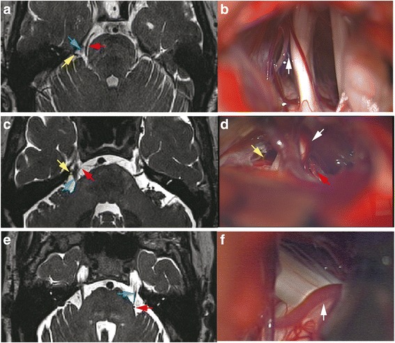
Fig. 2.

Routine MRI displaying NVC may have different intra-operative findings. a The superior cerebellar artery (SCA, red arrow) and a vessel (yellow arrow) located at the lateral of TGN (blue arrow) were the suspected vessels of NVC. b Intra-operative image showed SCA was close to but not in touch with TGN. A trivial vessel (vein) crawled on TGN. The white arrow points to the gap of the SCA and TGN. c MRI revealed the SCA (red arrow) and another vessel (yellow arrow) each formed a loop conflicting TGN (blue arrow) from inner side and outside. d Intra-operative image showed both vessels did not compress TGN (correspondingly marked with red and yellow arrow). A vein (white arrow) compressed the TGN from lower, outside at REZ to the TGN surface. e A very tiny vessel was displayed by pre-operative MRI at the outside of REZ of left TGN (blue arrow) which was easily neglected (red arrow). f An artery was found compressing the REZ of left TGN during surgery (white arrow)
