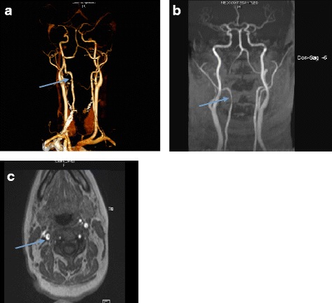
Fig. 1.

a CTA Neck : Focal stenosis with thickened wall and an intramural thrombus of left vertebral artery at the level of C3 (arrow). b and c A MRI coronal (b) and horizontal (c) view of the vertebral arteries confirms a 1.1 cm C2-C3 right vertebral artery dissection with no evidence of posterior circulation cerebral infarct (arrow)
