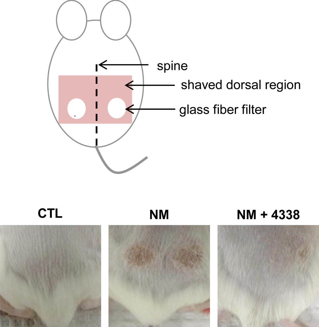
Fig. 2. Modified dorsal skin patch model.
The dorsal skin of CD-1 mice was shaved and 6 mm glass fiber filter discs placed on the lumbar region of the skin equal distant from the spine (upper panel). Twenty microliters of a 1 M solution of NM in 20% deionized water/80% acetone (v/v) or vehicle control was applied to the filters which were then covered with PARAFILM®. After 6 min, the filter discs were removed and the skin analyzed for tissue damage 1–5 days post exposure. Mice were treated with 4338 four times per day beginning 1 h post NM exposure. The lower panel shows the skin from control (CTL), NM and NM + 4338 treated skin 3 days post NM.

