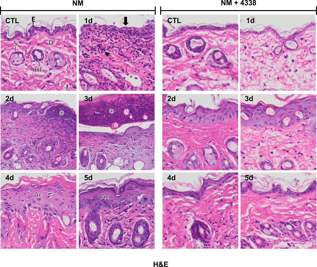
Fig. 3. Hematoxylin and eosin staining of mouse skin following NM exposure.
Histological sections, prepared from control (CTL) mouse skin and mouse skin 1–5 days post NM exposure, were stained with hematoxylin and eosin, which stains nuclei blue/black, and keratin and cytoplasm red. One representative section from 3 mice/treatment group is shown (original magnification, × 400). E, epidermis; D, dermis; S, sebaceous gland; HF, hair follicle; asterisk, eschar, Ac, acanthosis. Black arrow, loss of stratum corneum; black arrowhead, inflammatory cell infiltrate. Left panels, mouse skin treated with NM; right panels, mouse skin treated with NM and 4338.

