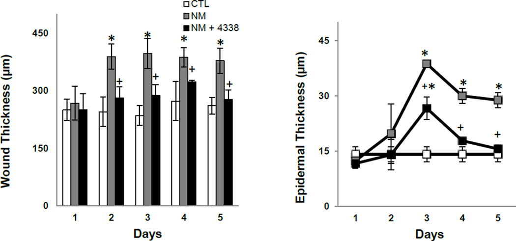
Fig. 4. Effects of NM on mouse skin wound and epidermal thickness.
Histological sections, prepared from control (CTL) mouse skin and mouse skin collected 1–5 days post NM, were stained with hematoxylin and eosin and wound (left panel) and epidermal (right panel) thickness assessed as described in the Materials and Methods Section. Each point represents the mean ± SE (n = 6). *Significantly different from control mouse skin (p≤0.05); +Significantly different from NM treated mouse skin.

