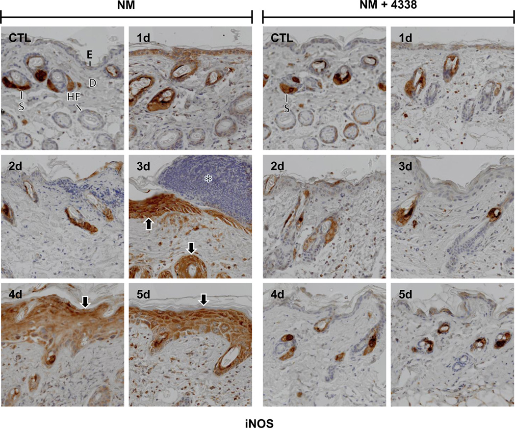
Fig. 7. Effects of NM on iNOS expression in mouse skin.
Histological sections, prepared from control (CTL) mouse skin and mouse skin 1–5 days post NM, were stained with an antibody to iNOS. Antibody binding was visualized using a Vectastain Elite ABC kit. One representative section from 3 mice/treatment group is shown (original magnification, × 400). E, epidermis; D, dermis; S, sebaceous gland; HF, hair follicle; asterisk, eschar. Black arrows, keratinocyte expression of iNOS. Left panels, mouse skin treated with NM; right panels, mouse skin treated with NM and 4338.

