Figure 4.

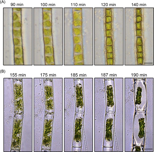
Light microscopic image series of the desiccation process in (A) Klebsormidium crenulatum at 78.3% RH, Scale bar 5 μm and (B) in Zygnema sp. at 56.4% RH, time in minutes (min) after the start of the desiccation experiment. Scale bar 20 μm.

Light microscopic image series of the desiccation process in (A) Klebsormidium crenulatum at 78.3% RH, Scale bar 5 μm and (B) in Zygnema sp. at 56.4% RH, time in minutes (min) after the start of the desiccation experiment. Scale bar 20 μm.