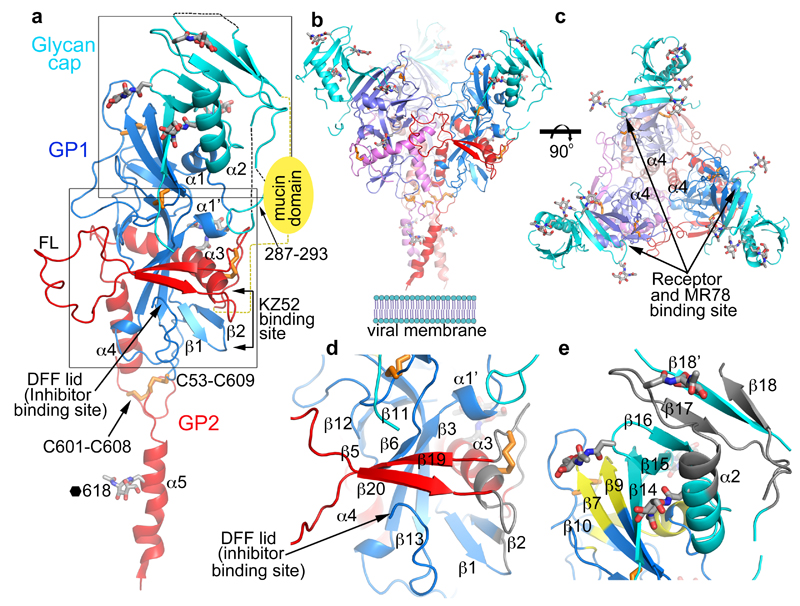
Figure 2. Overall structure.
a, Cartoon diagram of EBOV GP monomer, GP1 blue, GP2 red and the glycan cap cyan. Secondary structural elements named as previously8. Disulphide bonds shown as orange sticks, glycans in grey. The mucin domain omitted in our construct is shown as a yellow oval. FL, fusion loop. The C-terminal inserted foldon trimerization domain is disordered. b, The biological trimer viewed perpendicular to the 3-fold axis with one monomer coloured as in a and the second and third faded for clarity. c, The trimer viewed along the 3-fold, towards the viral membrane. d, Close up of the inhibitor binding site. e, Close up of the glycan cap and receptor binding site. Areas shown in d and e are indicated in panel a. In d and e antigenic sites are coloured grey and the receptor binding site yellow.

